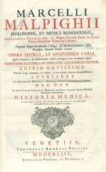
Marcello Malpighi (1628 - 1694) — Auction price
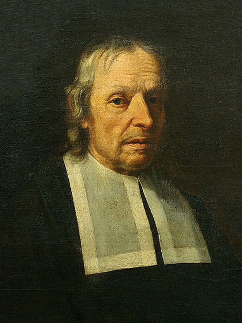
Marcello Malpighi was an Italian biologist, anatomist and physician, professor of logic, theoretical and practical medicine, and a member of the Royal Society of London.
After graduating from the University of Bologna with the degree of Doctor of Medicine and Philosophy, Malpighi soon took up a professorship there, then taught at the universities of Pisa and Messina. At the same time as teaching, he conducted biological research with his microscopes, which was an innovation in those days. In 1661, he identified and described the pulmonary and capillary network connecting small arteries to small veins, one of the most important discoveries in the history of science. He also isolated taste buds and regarded them as nerve endings, described the minute structure of the brain, the optic nerve, and in 1666 was the first to see red blood cells and attribute to them the color of blood. His treatise De polypo cordis (1666) explained the composition of blood and how it coagulates.
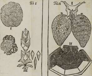
During his medical practice, Malpighi studied microscopic sections of the liver, brain, spleen, kidneys, and the bone and deep layers of skin that now bear his name. In his landmark 1673 work on the embryology of the chicken, the scientist concluded that the embryo forms in the egg after fertilization. In 1675-79 he also made extensive comparative studies of the microscopic anatomy of several different plants and saw analogies between plant and animal organisms. The Royal Society of London published two volumes of his botanical and zoological works in 1675 and 1679. His Anatome Plantarum is richly decorated with engravings by Robert White.
After his house was burned and looted by his adversaries, in 1691 Pope Innocent XII invited him to Rome as papal personal physician, which was a great honor.
Malpighi can be considered the first histologist. For almost 40 years he used the microscope to describe the main types of plant and animal structures and thus marked for future generations of biologists the main directions of research in botany, embryology, human anatomy and pathology. The conflict between ancient ideas and modern discoveries continued throughout the seventeenth century. Malpighi was convinced that microscopic anatomy, by showing the minute structure of living things, questioned the value of the old medicine. He laid the anatomical foundation for the subsequent understanding of human physiological exchanges.


